Cytological analysis in gynecology is often carried out. It involves the study of a taken tissue sample for the usefulness of the cell structure. The type of diagnostics allows relatively quickly and with high accuracy to determine the presence of inflammation, cancer or a precancerous condition of the female genital organs.
Cytological examination, as well as taking material for it, does not involve dissection or puncture of tissues. In order for the answer to be informative, it is necessary to properly prepare for the procedure for taking the biomaterial.
How is the analysis done
A smear is taken during an examination by a gynecologist for the purpose of subsequent cytological examination (PAP test, Papanicolaou analysis). It is a biological material scraped from the mucous membrane of the cervix. Subsequently, the sample is carefully examined using a microscope. The action allows you to quickly, but reliably detect the beginning of cellular changes. In 9 out of 10 cases, they unmistakably indicate the development of cancer or a high predisposition to it.
The cytological examination of the smear is aimed at achieving two goals at the same time. The first is the detection of oncological neoplasms at the initial stage of their development. The second goal is to establish the microflora of the vagina, to identify its violations.
Features of a cytological study
- Thanks to the study, it is possible to identify 5 types of cellular changes.
- The attending gynecologist is engaged in deciphering the answer of the analysis. The results of the study are interpreted separately from the woman's complaints and the results of other types of diagnostics. But the listed criteria are taken into account when prescribing treatment.
- The response of a cytological study is especially important when biopsy is contraindicated.
- The method helps to quickly examine a large number of patients belonging to the so-called risk group for the formation of malignant tumors of the cervix.
- A cytological study is characterized by technical simplicity and affordability - every woman can and should undergo an analysis. The reliability of this type of diagnosis is not in doubt. Therefore, cytological examination has been introduced into gynecological practice and has been successfully used for several years.
In the practice of gynecologists, an alternative definition of this term is also used - “histological smear”.

Who needs to be analyzed?
A smear for cytology should complement the laboratory diagnosis of the condition of women of different ages. For example, before reaching the age of 45, it is important to take it once a year.
Given the increased predisposition to oncological neoplasms, women over 45 years of age should undergo such a diagnosis once every six months.
There are a number of indications, in the presence of which it is necessary to pass a smear for a cytological examination. These factors include:
- failure in the menstrual cycle,
- inflammation inside the cervical canal, cervix,
- infertility, frequent miscarriages, fetal anomalies in the past and other problems associated with the inferiority of the reproductive function,
- preparation for implantation of the intrauterine device.
- taking hormonal drugs
- diabetes mellitus, regardless of type,
- moderate to severe obesity
- confirmed presence of genital warts, infection with genital diseases,
- promiscuity,
- pregnancy planning.
Analysis of the species under consideration is one of the mandatory diagnostic steps in preparation for surgery.
There is a small list of contraindications: the period of menstruation, the presence of active inflammation in the vagina or cervix. Restrictions are associated with leukocytosis - an increase in the level of leukocytes. The phenomenon occurs whenever there is inflammation in the body.
An increase in white cells will prevent the visualization of atypical cells - the laboratory assistant may not recognize them, provide incorrect information. Therefore, a cytology test for inflammation and menstruation is not recommended.
How to prepare for the study
The task of the study is to confirm or refute a precancerous condition. It is also possible to identify a high predisposition to the oncological process. This is an important analysis, on the result of which further tactics depend. In some clinical cases, the patient is prescribed therapy, sometimes with the use of minimally invasive procedures. To get a reliable answer, on the eve of the analysis, the patient must follow certain measures:
- do not flush the genital tract with a syringe at least 3 days before the planned visit to the doctor,
- do not urinate 3 hours before the manipulation,
- refuse intimacy for 2-3 days before taking a smear.
In the presence of an inflammatory process with an abundance of vaginal secretion, it is first important to eliminate the pathology, and then undergo a control diagnosis. If, according to its results and in the absence of clinical manifestations, recovery has occurred, a cytological analysis can be performed. A similar scheme applies to the fact of menstruation - initially you should wait until it ends, and only then visit a doctor. A priori, in each case, on the eve of the study, it is important to conduct thorough intimate hygiene.
How to take a smear for analysis
 Cytological smear - Pap smear
 The material is taken only by the gynecologist during the traditional examination of the patient on the chair.
By manipulating the mirrors, the specialist examines the condition of the vagina, cervical canal, mucous membrane of the cervix.
Assesses the color and density of tissues, the presence of neoplasms and secretions (if present). The material is taken from 3 sites: from the vagina, cervical canal, uterine pharynx.
The action is painless, it is performed using a special atraumatic probe or brush (they are always supplied with a gynecological examination kit).
The procedure takes no more than 15 minutes.
The taken material is transferred and then distributed on a glass slide. Having slightly dried the sample in air, the smear is transferred to the laboratory. There is staining with reagents (these are special medical solutions), then there is a study under a microscope.
During this activity, the following criteria are evaluated:
- size, structure of cells,
- how many cells are localized in a certain area unit,
- features of their localization to each other,
- shape of the epithelial tissue
- the presence of pathology, the presence of deviations from the norm.
After the procedure, you can continue your daily activities. Unpleasant sensations should not be normal. If the manipulation is carried out by an inexperienced gynecologist, slight damage to the walls of the genital tract is possible. Then 1-2 days after taking a smear, leucorrhoea, slightly stained with blood, is observed. A similar complication is observed in cases where the patient's mucous membrane is enriched with a dense capillary network. Especially if it is close to the surface of the tissues. Then the blood vessels react even to minor interventions with brittleness and, as a result, light bruising.

Deciphering the answer of the analysis
With the full health of a woman, the cervix is lined with cylindrical epithelium, and the vagina consists of flat cells. The natural microflora of the vagina is represented by rods. Also, the analysis response displays indicators of the functional activity of the ovaries. Additionally, the result contains information about the level of leukocytes (their increased concentration confirms the presence of inflammation in the body).
Deciphering the Pap test
Taking into account his condition, the material taken from the walls of the vagina is classified as follows (Papanicolaou method):
- Class 1. There are no signs of pathological processes in the studied material sample. The size, shape, mutual arrangement of cells correspond to the norm.
- Class 2. Signs of inflammation and vaginosis were found in the material sample. The answer of the analysis serves as the basis for additional diagnostics, the need for therapy is not ruled out.
- Class 3. The laboratory assistant identifies units of cells with a disturbed structure of the nucleus and cytoplasm. The response of the analysis serves as the basis for repeating the procedure.
- Class 4. A precancerous condition is established. The material contains cells with malignantly altered nuclei, chromatins, and cytoplasms.
- Class 5. Contains a large amount of atypical cells - has arisen malignant neoplasm early stage.
Sometimes cytological diagnostics is combined with a biopsy - a method of taking a piece of tissue from an organ that is suspected of having a predisposition to cancer. The procedure is painful and is performed only after anesthesia has been performed.
The purpose of the method is to clarify the previously obtained information. In particular, if a precancerous condition or the actual tumor process of malignant origin is established on the basis of cytology.

Deciphering according to the Betsed method
Using this method, the doctor will decipher the answer of the analysis of the material, which is taken during examination from the cervical canal. The action takes place with the obligatory consideration of the localization of cells and changes within the nucleus.
Only one of the possible answers of laboratory diagnostics is received. Marking "norm" indicates the absence of disease-causing changes. Vaginosis and koilocytosis are codenamed HPV. If cervical dysplasia is detected, the CIN I, CIN II or CIN III code is present in the analysis response (depending on the established degree). A malignant tumor of the cervix is noted as Carcinoma (pax).

Common terminology in cytology response
In the process of gynecological examination and receiving answers cytological analysis, use the following terminology:
- CBO - the state of the cervix without deviations from the norm.
- Cytogram of inflammation - there is cervicitis (inflammatory process of the mucous membrane of the cervical canal).
- Leukocyte infiltration - an increase in the concentration of white cells, presumably vaginosis, exocervitis or endocervitis is present.
- Koilocytes are cells characteristic of the course of HPV (human papillomavirus).
- Proliferation refers to the process by which cells divide at an accelerated rate. A similar reaction is characteristic of intrauterine inflammation.
- Leukoplakia - the material contains signs of pathology. Despite intracellular changes, there is no development of cancer.
- Metaplasia is the process of replacement of cells of one type by another. It is not a pathology if a woman underwent therapy for non-oncological uterine changes during menopause. The process accompanies the state of the body of patients who are in a state of menopause for more than 6 years.
- Dysplasia is directly a precancerous phenomenon.
If the received answer raises doubts in the doctor, the specialist prescribes an even more thorough examination to clarify the clinical situation. A woman takes a smear in order to clarify the nature of the flora, undergoes an analysis to determine venereal diseases.
When a material sample contains abnormal cells, laboratory technicians use the following abbreviations:
- ASC-US - for unknown reasons, there is a change in the structure of the squamous epithelium. Often detected in middle-aged women, in the premenopausal period, since it is characterized by hormonal instability.
- AGC - a change in the structure of cylindrical cells (it happens with vaginosis, it is typical for other inflammatory disorders). The cause-and-effect relationship is clarified due to additional diagnostic measures.
- L-SIL - the presence of a small number of degenerated cells of non-cancerous origin. The patient will undergo a colposcopy procedure, supplemented by a biopsy.
- ASC-H - intracellular changes indicating the presence of a precancerous pathology or a tumor process that has already begun.
- HSIL - smear contains flat cells, serves as a precancerous condition. Carry out immediate therapeutic measures, thereby preventing the process of degeneration of pathology into cancer.
- AIS - detection of cylindrical cancer cells. The patient is treated urgently. If the presence of degenerated cells is laboratory proven, this fact must be written in the analysis response, specifying the type of changes.
When the decoding of the analysis response does not contain specific markings, the smear indicates the normal state of the female body. Establishing the nature of the violation, the doctor focuses on the results of different types of diagnostics.
How long will it take to get the result
The answer is received after 2-5 days. Sometimes this period is affected by the workload of laboratory assistants.
It is important for the patient to understand that the process of developing an oncological neoplasm does not occur instantly. It is quite possible to recognize atypical cells at the stage of their degeneration. To do this, you should undergo an analysis systematically, visit a doctor without passes.
From the moment the transformation of physiological cells begins to the appearance of the first signs of oncology, a significant period of time passes. And this period is enough to get the answer of the analysis passed, to recognize cervical cancer (if its development is still relevant) and to begin the therapeutic effect.
Today, cytological examination is recognized as the best option for the timely detection of a malignant tumor of the cervix.
30 ratings, average: 4,83
out of 5
Content
A cytology smear, traditional or liquid, is one of the most important links in the screening diagnosis of precancer and cervical cancer. The introduction of the method into cytological screening programs has reduced mortality from cervical cancer in many countries.
Due to the high level of information content, simplicity and ease of implementation, safety for the patient, a cytology smear or PAP test is widely used in mass preventive examinations, and has established itself as an indispensable way to detect patients with underlying and precancerous diseases of the cervix.
A smear for cytology allows you to identify patients in the pre-symptomatic phase of cancer or dysplasia, apply sparing methods of treatment in a short time, and not disrupt reproductive plans.
A smear for cytology has a low sensitivity, therefore ideal option diagnostic examination of the patient for background and precancerous pathologies is a combination of several methods:
- colposcopy;
- biopsies with histological examination of tissue;
- PCR for human papillomaviruses or its improved version Digene test.
The complex technique provides 100% accuracy of results with high-quality material taking and its correct assessment.
It is known that the cause of a malignant tumor of the cervix is at least 15 types of human papillomavirus, and two of them - 16 and 18 - initiate a tumor in 70% of cases. Therefore, the diagnosis of diseases of the cervical region should also include a smear from the cervical canal for HPV. If a virus is detected in a smear, appropriate therapy is carried out, which significantly reduces the risk of developing precancerous pathologies.
The advantage of a combined examination, which includes not only a smear for cytology, but also an HPV test, is the possibility of preventing the formation of adenocarcinoma, a malignant tumor that is not detected when performing a cytological method.
Timing and indications for the test
The first smear for cytology, as a rule, is taken by young women at the age of 18. However, such an analysis is often started at the age of 21. The frequency and timing of the smear does not depend on the intensity of the girl's sexual life.
Smear frequency:
- from 18 years (21 years) to 64 years, a smear should be performed once a year;
- patients over 65 years of age perform the analysis once every 3 years, provided there were no atypical cells earlier;
- once every six months, a smear should be performed in case of menstrual irregularities, the presence of HPV, grade 1 dysplasia and ectopia complicated by infections, while monitoring the therapy of cervical pathologies.
The most appropriate time for performing cytology is the middle of the menstrual cycle. The period before menstruation, as well as after them, is undesirable for a smear due to specific hormonal changes in the cervix.
The completion time depends on the workload of the laboratory, as well as on the type of structure: public or private. As a rule, in public institutions, the result of cytology is ready in 7-14 days, and in private institutions - after 1-3 days.
Features of the smear procedure for cytology
The material is considered adequate for research if the cytologist detects in it the cells of the cervical canal, the vaginal part of the cervix and the transition zone of transformation: squamous epithelium, intermediate and metaplastic, cylindrical in the endocervix, single erythrocytes.
It is mandatory to obtain a smear from the transformation zone - the area most susceptible to malignant changes. The smear is considered inadequate in the absence of cells of a cylindrical epithelium, a large number of erythrocytes, leukocytes, with a meager number of cells. If the smear for cytology is too thick or thin, the effectiveness is sharply reduced.
The procedure for taking a smear consists of several stages and has features.
- The gynecologist performs two-handed palpation of the uterus, cervix and ovaries.
- Colposcopy should be performed for the most accurate cytology result. With multiple magnification, the cervix is examined for pathological inclusions. After that, the doctor treats the surface with a solution of vinegar and evaluates the result. In the presence of persistent white staining (acetowhite epithelium), a smear for cytology is taken aimingly from these places. They also act when the neck is lubricated with iodine and there is no brown staining (iodine-negative reaction). A smear is taken from unstained places, since this reaction of the integumentary epithelium is considered a pathology.
- Instruments must be dry and sterile, preferably individually wrapped. Water, disinfectant solutions can destroy cell samples, which will adversely affect the result of cytology. When taking a smear, specialists use special tools: Cervix-Brash, Papette cytobrushes, modified Eyre spatulas.
- Taking a smear for research is taken from the surface of the cervix and the visible part of the canal, including the transformation zone, which is clearly defined during colposcopy. In addition, using a spatula, a smear is taken from the outer surface, a scraping from the cervical canal is performed with a cytobrush.
- The selected material is either applied to glass (in classical cytology) or immersed in a transport liquid (liquid-based cytology).
- Test tubes and glasses are marked.
During manipulation, a woman may feel slight discomfort. After a smear for cytology, brown discharge from the genital tract is noted throughout the day.
Pregnant women over 22 weeks gestation Cytology is performed strictly according to indications, as the procedure can cause complications.

Goals and objectives
A smear for cytology allows you to identify a precancerous pathology of the cervix - epithelial dysplasia, in which the risk of developing preinvasive cancer is 20 times higher. The transition of stages 2 and 3 of dysplasia to preinvasive cancer (in situ) lasts from 3 to 8 years, and after 10-15 years, microinvasive cervical cancer develops.
The most common pathologies detected by smear cytology are:
- ectopic columnar epithelium;
- hyperkeratosis, parakeratosis - violations of keratinization of the squamous epithelium;
- glandular hyperplasia;
- chronic cervicitis;
- various stages of dysplasia and cancer.
Cytology allows you to identify a dysplastic process in the epithelium of the cervix, which prevents the formation of cancer with timely treatment. Cervical dysplasia is asymptomatic at stages 1 and 2, so an annual smear for cytology contributes to the early detection of a dangerous pathology.
Stage 3 dysplasia and pre-invasive cancer are most commonly reported in women who do not visit a gynecologist for 5 consecutive years and do not perform a cytology smear.
When reviewing cytology smear results, it is important to evaluate all diagnostic studies, in particular histology or biopsy.
Liquid cytology method
The traditional smear for cytology has several limitations that lead to a false negative result (within 2-50%), while the main source of errors in screening and evaluation of results is poor-quality collection, processing of the obtained material and qualification of the laboratory assistant.
Therefore, the classical smear was replaced by cytology new method- liquid cytology. This method was developed in 1996 in the USA. Its essence lies in the immersion of the material from the surface of the cervix and cervical canal not onto a glass slide, but into a liquid accumulation medium. A single-layer preparation is prepared from the resulting suspension using an automatic device, which leads to an increase in the efficiency of evaluating the results. When performing a classic smear for cytology, the preparation consists of several layers of cells, which makes it difficult to correctly assess, the true picture may be distorted.
An important feature of the method of liquid cytology is the collection of material obtained by smear from the cervix in a special environment that contributes to the preservation of cells, which increases the effectiveness of the study. The structure of cellular elements and their immunohistological characteristics are completely preserved. This allows you to process the resulting smear with special reagents and carry out immunocytochemical reactions and hybridization.
After extracting the cells from the liquid medium, staining is carried out. Staining is performed in the same way as in a traditional smear for cytology, for example, according to the Pappenheim method.
Decryption
For the development of many pathological phenomena in the area of the integumentary epithelium of the cervix, the peculiarity of the anatomy of the organ is of decisive importance. In particular, the relationship between the layers of the epithelium of the cervical canal and the vaginal portion of the cervix plays a role.
As a rule, all atypical processes, and then malignancy, occur in the area of the transition of the stratified squamous epithelium, which forms the surface of the neck, into a cylindrical, lining the channel from the inside. The cylindrical epithelium is otherwise called prismatic or glandular, since its main function is to produce a protective mucous secretion to form a plug. The area of transition from one type of tissue to another is called the transition zone or the transformation zone. A smear for cytology should be taken, including this area.
The transformation zone can be located in various places:
- on the surface of the cervix in young women, as well as during pregnancy and after childbirth;
- at the entrance to the cervical canal- in women of the reproductive phase;
- deep inside the cervical canal- in women of mature age and in menopause.
In 96% of cases, pathological processes occur precisely in the transitional area, and in the rest - in the area of the cervical canal.
After staining the smear, the cytologist carefully examines the sample under a microscope. In this case, the specialist evaluates:
- the type of cells identified and their affiliation (squamous, cylindrical, intermediate epithelium and metaplastic);
- sizes of cellular elements;
- maturity;
- state of the cytoplasm and nucleus;
- the ratio of the size of the cytoplasm to the nucleus;
- division intensity.
To decipher a smear for cytology, various classifications are used for the purpose of unification. The evaluation of results according to the Papanicolaou system is widely used.
- The first class smear means the norm.
- The second class of cytology is characterized by inflammatory changes.
- The third class is described by the presence in a smear for cytology of cells containing atypical nuclei, cytoplasm - dysplasia is not excluded.
- The fourth class means the presence of atypical cells that do not exclude cancer.
- For the fifth class, large numbers of cancer cells are inherent.
One of the most popular is the Bethesda system. This classification implies 3 types of smears.
- NILM is the norm, namely the absence of intraepithelial malignant changes in the smear. Cytology has a normal result in terms of dysplasia and cancer, but does not exclude inflammatory, atrophic and other changes, for example, the result may indicate the presence of a large number of leukocytes, Trichomonas, yeast, bacteria (cocci), viral changes in epithelial cells.
- ASCUS - a cytology smear is indeterminate, there is atypia of unknown origin.
- ASC-H - atypical squamous cells are present in the smear, and this finding does not exclude a high degree of dysplasia.
- LSIL - changes detected in the smear indicate a low degree of intraepithelial changes associated with human papillomavirus - dysplasia grade 1 (CIN I).
- HSIL - means the presence of high-risk changes or 2, 3 levels of dysplasia (CIN II, CIN III). In addition, the smear result does not rule out preinvasive (in situ) or microinvasive cancer.
In addition to dysplasia, a cytology smear detects cancerous changes, which are designated AGC, AGUS, cancer in situ, squamous cell carcinoma (High-Grade SIL), or glandular (adenocarcinoma).
For suspicious cytology smear results a woman is given a biopsy that confirms or rules out cancer.
After receiving the results of histology, the doctor determines the tactics of further actions. Additional tests and examinations are carried out depending on individual situations:
- multislice computed tomography;
- MRI with contrast;
- bone scintiography;
- angiography;
- Ultrasound of the pelvis and abdominal cavity;
- x-ray studies;
- blood tests for tumor markers;
- diagnostic laparoscopy.
When confirming the initial stages of the tumor, or rather preinvasive cancer or stage 3 dysplasia, conization is performed with the capture of healthy tissues. Methods such as radio wave conization, laser, electric loop are used. The removed cone is examined histologically to assess the quality and completeness of the excision. A woman is prescribed a course of immunomodulators and is observed for the purpose of a dynamic assessment of the state of the cervix. If microinvasive or stage 1 cancer is detected, hysterectomy is performed. The second and subsequent stages require not only the removal of the organ, but also chemotherapy, radiation therapy.
Many countries have uniform cytological screening programs. In Russia, at the legislative level, it is customary to examine all women who have reached the age of 18, with a smear for cytology from the cervix and cervical canal.

In the work of gynecologists assisting in the diagnosis of cervical pathologies, there should be no formalism, sampling for analysis should be carried out taking into account the identified external changes on the surface of the organ, and the result should be evaluated in conjunction with all diagnostic methods. The choice of a clinic for cervical cancer screening should be treated scrupulously, as it is not uncommon for situations when after the tests it is not possible to detect initial changes.

 The complexity of diagnosing diseases of the genital organs in women often leads to a loss of time required for successful treatment. Often, the development of cancer cells is asymptomatic, and only a special examination can reveal a dangerous disease. A smear for cytology or a pap test in women allows you to identify cancer at an early stage and start treatment on time.
A cytological examination of the tissues of the cervix in women allows you to determine 5 types of changes in the cells. A smear for cytology is inexpensive and effective method diagnostics, used in medicine for more than 50 years. It is recommended to conduct a study for all women without exception aged 21 to 65 years at least 1 time per year. Deciphering the Pap test gives a complete picture of the presence or absence of any abnormalities.
 A cytological smear (Pap test, Pap smear, smear for oncocytology) is performed during a gynecological examination. The doctor uses a mirror to examine the vagina, the entrance to the cervical canal and the cervical mucosa. If there is a suspicion of an anomaly, cells are taken with a special brush from 3 areas: from the walls of the vagina, the cervical canal, the entrance of the cervix. The procedure is comfortable, painless and does not require special preparation.
The mucus is applied evenly to the glass slide, dried and sent to the laboratory.
The laboratory assistant with the help of reagents stains the smear, examines it through a microscope. This method determines the indicators:
- cell structure;
- cell size;
- the shape of the epithelium;
- mutual arrangement;
- the number of cells per unit area;
- pathological changes in the structure of cells.
 A cytology smear allows you to identify most inflammatory diseases, precancerous pathologies of the epithelium (dysplasia), and malignant tumors. After taking a smear, spotting is often observed for 2-3 days, which is normal. Extremely rare - severe bleeding, abdominal pain, chills, fever. In this case, an urgent examination by a gynecologist is required.
When is an analysis ordered?
 Ideally, every woman can undergo a cytology test regularly, without special instructions from a doctor. A routine gynecological examination can reveal the presence of inflammation of the cervix and cervical canal. A cytological smear is only a method of confirming the diagnosis. Therefore, it is better to adhere to the recommendations of the gynecologist - if there are no indications for analysis, then you should not worry ahead of time.
However, a cytology study should be carried out for women under the age of 40 - once a year, older women - 2 times a year and more often. Cases in which a cytological examination is prescribed without fail:
- with menstrual irregularities;
- in inflammatory processes of the cervical canal, cervix, etc., especially chronic ones;
- in violation of reproductive function;
- during pregnancy planning;
- before surgery and other medical procedures;
- before installing an intrauterine device;
- taking hormone-containing drugs;
- obesity 2, 3 degrees;
- diabetes;
- the presence in the body of the papilloma virus, genital herpes;
- active sex life of a woman with frequent change of partners.
How to prepare for analysis
 To ensure the maximum degree of purity of the smear, you should follow the rules before going to the gynecologist:
- Do not use local drugs (vaginal tampons, suppositories, ointments).
- Do not douche.
- Wait until the end of the month.
- In inflammatory diseases with abundant secretion, a general treatment should first be carried out. After a control smear confirming recovery, you can proceed to the analysis for cytology.
- You can not urinate 3 hours before the cytological analysis.
- It is better to refrain from sexual intercourse 2 days before taking the secret.
Compliance with these rules will avoid unnecessary anxiety and repeated visits to the doctor.
If the doctor prescribed a smear for oncocytology, this does not mean that the doctor has made a terrible diagnosis and is waiting for its confirmation.
Remember: prevention is better than cure.
What can a cytological analysis reveal?
How to decipher a smear for oncocytology? The interpretation of the data obtained in the laboratory is understandable only to the doctor. And not always the gynecologist gives a detailed picture of the disease, not wanting to waste time on explanations. 
In the process of research, you can get 5 results:

It is important to remember that a smear shows only the degree of cell change, the presence of inflammation, infections, but does not determine exactly the cause that causes them.
On the basis of only a study on cytology, the gynecologist does not make a diagnosis, for this a comparison with other analyzes is necessary.
2, 3, 4 type of changes revealed in the study of cytology, may be a sign of diseases:
- ectopia (erosion) of the cervix;
- papillomavirus infection;
- herpes genital;
- parakeratosis of the cervix;
- bacterial vaginitis;
- cercivit;
- vaginal candidiasis, etc.
Result interpretation
Deciphering the results of the analysis for the doctor is a simple matter, for the patient it is incomprehensible letters and terms.

If atypical cells are found in the smear, the laboratory assistant will write about this in the conclusion, and also determine the type of changes. Therefore, if the transcript of a smear for cytology does not contain special notes, then most likely no pathologies were found. 
The time for a smear for cytology is from 1 to 5 days. Pathological changes in the cells of the cervical canal and cervix on the way to the diagnosis of "cancer" go through several stages, and not in 1-2 days. Cytological examination allows identifying atypical cells at the initial stage and starting treatment, which in most cases leads to complete recovery. Therefore, cytological examination has been widely introduced into medical practice as a quick, painless and inexpensive way to diagnose cancer cells at an early stage.
Cytology of the cervix is a laboratory study that determines the cellular structure of the cervix uteri (cervix), as well as the cells of the cervical canal. One of the variants of cervical cytology was first used at the beginning of the 20th century by a Greek scientist Papanikolaou for the purpose of early detection and prevention of oncological diseases in gynecology.
Since then, the PAP test has been used everywhere along with a new method - ThinPrep (liquid cytology), which allows timely detection and effective treatment of latent chronic and precancerous diseases of the cervix.
, , , ,
Indications for carrying out
The main task that cytology of the cervix solves is the prevention of the oncological process. Early detection of atypical cells makes it possible to stop cervical cancer (cervical cancer) in time, which, according to statistics, is in third place among the most common oncopathologies in women. The risk of an asymptomatic course of the disease is high, therefore the main objective procedures are the timely detection of precancerous changes in cells. This makes it possible not only to increase the percentage and survival time, but also a confident hope for a successful cure. The PAP method in the practice of gynecology is considered one of the fastest and most accurate for obtaining data on the presence or absence of the initial stages of oncological processes, cancers, or background pathologies of non-tumor etiology that can provoke life-threatening cancer. Many doctors call cervical cytology the "gold standard" for diagnosing and screening for such intraepithelial changes and conditions:
- Various cellular anomalies of high or low degree.
- The presence or absence of pathogenic microflora for the diagnosis of the state of the mucous membrane of the cervix.
- Violations of the normal rhythm of the menstrual cycle (frequency of the cycle, its delay or absence).
- All types of diseases of viral origin - HPV (human papillomavirus) - warts, Herpes simplex genital (herpes).
- Infertility (infertility).
- Defects of the mucous membrane of the cervix (all types of erosion).
- A long cycle of therapy with hormonal drugs.
- Abnormal vaginal discharge, including blood, especially after sexual intercourse.
Cervical cytology is needed as a screening test if:
- Planned pregnancy
- Births occur several times in a row (for example, 3-4 times within 4 years).
- The first birth occurred at an early age (up to 18 years).
- A woman often changes sexual partners.
- Planned contraception in the format of an intrauterine device.
- Previously, cytology has never been performed, or the woman has not been examined for the last 3 years.
- The last sampling of tissue material does not meet the standards or identified changes in the cells.
- Examination of the cervix with a mirror shows visible pathological changes (color, structure).
- The woman was diagnosed with diseases associated with immunodeficiency (HIV).
- Family history is aggravated by oncological diseases (one of the direct relatives was ill or is currently ill with oncological disease).
In general, an annual screening cytology examination should be mandatory for every woman who has reached the age of majority. If the cytology of the cervix shows pronounced clinical deviations of the cellular material, the examination should be carried out more often - at least 2 times a year or in the mode of a comprehensive therapeutic plan.
Preparation
Preparation, which involves the cytology of the cervix, does not belong to the category of complex. The procedure itself takes place very quickly, with virtually no discomfort and discomfort, and the preparation consists in the following steps:
- Exclude sanitation (hygienic procedures) in the form of douching
- A few days before the procedure (2-3 days) you should adhere to the regimen of abstinence in intimate life
- Do not use hygienic tampons, tablets, liquids, creams, suppositories and vaginal gels
- 2 hours before the test, a woman should refrain from urinating
In order for cervical cytology to show a reliable result, it is important to know the following information:
- Pap test and menstrual cycle are not compatible. The procedure is carried out 3-5 days before the onset of menstruation or after them, it is best to do this on the 10-12th day of the cycle.
- Cytology will not be accurate in any infectious disease, especially in the acute phase, as a rule, the analysis is carried out after basic treatment. An exception is the need to obtain a cito result, in which case, cervical cytology is performed twice - during illness and after 2 months for cytological control.
- A smear during intravaginal therapy will not be informative, it should be done 5-7 days after the end of treatment.
- It is not recommended to take tissue material during inflammatory processes in the vagina, the indicators of which may be discharge, itching, burning.
Clarifications of the conditions for preparing for the procedure can be obtained from the attending gynecologist.
Technique
Cytology of the cervix is considered a mandatory procedure for a comprehensive gynecological examination. This is a standard that allows you to timely identify the risks of many diseases, including cancer.
The gynecologist makes a scraping from the outer part of the cervix (exocervix), as well as from the surface of the vaginal mucosa with a special spatula (Ayre spatula). The cells of the cervical canal (cervical canal) are taken with the help of an endobrush - a cervical probe, which makes it possible to obtain a sufficient amount of informative material.

Also, the doctor can use the following tools to collect cellular material:
- For a smear of the posterior fornix of the neck - Eyre's spatula.
- Aspiration tool - spirette, for sampling endocervical material.
- The screen is also designed to take biological material from the cervical canal.
- Cervex-Brush is a sterile medical brush for taking a smear from the T-zone (the surface of the cervix and cervical canal).
- Standard tweezers.
- Gynecological mirror.
- Cervical double-sided spoon (Volkmann's spoon) for a smear on the microflora of the mucous membrane of the cervical canal and the determination of infectious diseases.
All modern instruments are sterile, most of them are used individually for each woman.
Cytology technique:
- Examination on a gynecological chair with the help of mirrors, at the same time, material is taken for cytology. The walls of the vagina are expanded, the smear procedure (scraping) can cause short-term mild discomfort, but most often the cytology is painless.
- Simultaneously with the scraping, material for bacteriological examination (microflora) is also taken. Together with cytology, colposcopy can be performed, but this happens only according to indications, usually this procedure is prescribed separately.
- Samples of the material are applied to a special glass, fixed, fixed and transferred to the laboratory assistant for accurate microscopic examination using staining. There is also a new method of cytology of the cervix - liquid (ThinPrep), in this case, the material is placed in a flask and also transferred to the laboratory.
Analysis labeling is very important. The laboratory should receive glass with the material and a special form, which indicates the following information:
- Glass number in accordance with the direction number of the form.
- The name of the medical facility where the procedure was performed.
- Date of analysis.
- Name of the patient.
- Patient's age.
- Numbers, dates of the last menstrual cycle.
- Provisional (clinical) diagnosis.
Cytology of the cervix does not take much time, the whole procedure lasts no more than 10-15 minutes.
What does the cytology of the cervix show?
The main indicators of cervical cytology are in a nutshell - a negative or positive result, in other words, the test shows whether there are signs of cancer, infectious, bacterial, viral pathology in the smear.
Let's take a closer look at what the analysis shows:
- A negative result means that the epithelial cells are not affected by pathological processes, pathogenic flora is not detected, the cell structure is not destroyed by viruses
- Positive indicators indicate that abnormal cells were found in the mucous membrane of the cervix, their structure and number go beyond the standard norms. Atypical cellular elements can be diverse in shape, type and size, respectively, the interpretation of the analysis is directly related to these criteria.
In modern gynecological practice, the classification of results from the author of the test, the Papanicolaou technique, is considered traditional for use. The PAP-smear test method involves a detailed description of the composition and structure of cells, dividing them into 5 categories:
- Stage I - no cellular changes were found, there is no atypia, which indicates a practically healthy state of the cervix. The cytological picture is within the normal range
- Stage II - the test found a number of abnormal cells with signs of inflammation, which can be considered a relative norm, given the prevalence of inflammatory processes in the genital area. As a rule, a woman is prescribed additional diagnostic procedures to clarify the cause, nature, stage and causative agent of inflammation.
- Stage III - analysis shows the presence of a small number of locally grouped atypical cells with abnormalities in the structure of the nucleus or cytoplasm. This is also not a direct indication of a threatening pathology, but it may be a signal of the risk of developing an oncological process. An additional procedure that refines the PAP test may be a morphological analysis of the tissue material of the cervix (histology). A biopsy helps eliminate risks or confirm the onset of a pathological process that can be stopped with timely treatment.
- Stage IV analysis shows a clear picture of malignant transformations of a small number of cells. As a rule, abnormal cells have too large a nuclear mass, cellular contents (cytoplasm), chromosomes also show signs of pathological changes. This stage indicates the possible onset of cancer (dysplasia). Additional examinations help to clarify the diagnosis - colposcopy, sampling of material for histology, and repeated cytology is also possible.
- Stage V-test determines a fairly large number of altered cells, which indicates the presence of an oncological process. In such cases, the results of cytology are the beginning of a comprehensive diagnostic process aimed at determining the type of oncology, its stage and the vector of therapeutic measures.
Deciphering the results
Only the attending physician can and should interpret the test results. Any information obtained in the bowels of the Internet can only be exploratory and primary. The following data is publicly available and will help reduce the anxiety of women who are afraid of incomprehensible numbers and characters in the analysis.
Cytology of the cervix according to statistics reveals the following:
- Every ninth test out of ten determines the norm, that is, the next cytology can be safely carried out in 1-2 years for the prevention and screening of the cervix. The analysis should be repeated regularly, despite a good result, since it cannot be 100% accurate and does not completely eliminate the risk of diseases.
- Every second out of a hundred smears is not fully informative due to the lack of biological or cellular material. In such cases, cervical cytology is repeated
- Every twentieth test shows the presence of atypical changes, but this does not at all indicate a developed oncological process. Rather, it is a signal and a reason for a more thorough examination and subsequent treatment.
- The test result may show poor performance, but this cannot be considered an unambiguous and final diagnosis. To clarify the nature of the pathological process and its prognosis, additional examinations are required, which are prescribed by the doctor.
Deciphering the results of the cytogram can be carried out by different methods. The Papanicolaou classifier is classic, but in modern gynecology it is also customary to take into account other options for interpreting the test, for example, the American Bethesda System.
Information on the state of the epithelial tissue of the cervix can be reflected in the following table:
|
Presence of benign changes |
Pathological changes in epithelial tissue: dysplasia, atypia |
Invasive cancer |
|
|
Squamous cell invasive cancer |
|
The patient needs additional examination, observation and complex treatment. Screening cytological control is also required. |
Additional diagnostic procedures needed |
Histological examination of the material is necessary, possibly, clarification of the diagnosis after surgery |
Also, you can be disturbed by the Latin abbreviation in the analysis form, although in fact everything is deciphered simply, according to the “territory” of the material sampling:
- The urethra is the letter U.
- Endocervix, cervical canal - C.
- The vagina is the letter V.
The attending physician will provide you with the rest of the information.
, , , , , , , , , ,
Norms of cytology of the cervix
The norm in the cytology of the cervix is the complete absence of atypical changes in the cellular material, as well as the healthy state of the microflora. "Good" cells must meet the norms according to morphological standards, that is, in size, content (structure) and shape. The cytogram describes in detail the material taken from the surface layer of the cervix and from the endocervix.
The norms of cytological examination look like this:
- Cytology of the cervix contains cells of a single-layer cylindrical epithelium. Alternatively, the test may show the presence of stratified epithelium (metaplastic), this is also considered normal if the smear was taken in the transition area into the vagina.
- Stratified epithelial cells in a smear from the area of the vaginal part of the cervix are also considered normal if no changes are found in them.
- Any deviations from the norm, changes in cell morphology are described in the conclusion and may be indicators of primary inflammatory processes that fit within the normal range.
- Deviations from the norms in the PAP test do not mean the presence of an oncological process, with the exception of the HSIL indicator. This indicates a high risk of developing cancer.
It should also be borne in mind that there are indicators indicating benign cell abnormalities. This, of course, is not the norm, but it cannot be a reason for increased anxiety about oncology.
The list of changes within the relative norm that cytology can show:
- Atypia of inflammatory etiology.
- Atypical changes provoked by the papilloma virus.
- Mixed atypical changes.
- Atypical deviations of an unclear nature that require clarification.
The processes in which there may be deviations from the normal boundaries include the following diseases:
- HPV is the human papillomavirus.
- Herpes.
- Pregnancy.
- Long-term use of drugs, especially antibiotics.
- The use of contraceptives (candles, tablets).
- Contraception with a spiral.
According to the classification of Trout and Papanicolaou, the first 2 out of five classes can be considered the norm of cervical cytology, namely:
- I - normal cytological picture
- II - ASC-US or presence of atypical cells, unspecified
Poor cytology of the cervix
What do bad Pap test results mean?
Poor cervical cytology does not necessarily indicate the last stage of cancer. An exact decoding of the study can only be given by a specialist, that is, a gynecologist. According to the generally accepted classification, poor cytology is pathological changes in the epithelial layer of the cervix and cervical canal.
According to the generally accepted methodology, changes are indicated as follows:
- 0 - the material for the test is unsatisfactory (poor quality, in small quantities, uninformative due to insufficient patient preparation).
- Grade 1 is the norm.
- Grade 2 - the presence of atypical deviations.
- Grade 3 - various degrees of dysplasia.
- Grade 4 - precancerous condition, initial stage.
- Grade 5 - invasive cancer.
Grade 5 is considered the most alarming of the above changes, but any form of dysplasia is also a dangerous signal. Consider the types of dysplasia in more detail:
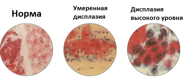
- A mild degree of dysplasia is a sign of an inflammatory process that has begun. It should not be skipped, since the inflammation can proceed almost asymptomatically and in the future go into a more severe form.
- Moderate dysplasia is already a threatening signal, indicating that the risk of developing an oncoprocess is very high.
- A severe degree of dysplasia is almost a precancerous condition.
Undiagnosed dysplasia, along with other provoking factors, can lead to a really dangerous condition - an oncoprocess.
Any woman, having learned that the cytology of the cervix shows a poor result, is afraid. Let's try to reduce its level a little with the help of informing.
What if the test showed the presence of obvious atypical cells?
First of all, do not panic, but listen and follow all the doctor's prescriptions. The gynecologist usually conducts a detailed consultation, both about additional diagnostic procedures, and about the prospect of treatment and the prognosis of the disease.
Usually the following events are appointed and held:
- Repeat cytology of the cervix.
- Biopsy (histological analysis of the tissue of the affected area of the cervix).
- Curettage of the epithelium of the cervix.
- An extended blood test.
- HPV test.
- If dysplasia is detected, it is treated (more often with the help of cauterization).
- If a concomitant viral infection is determined, not only the woman is treated, but also her sexual partner.
Timely diagnosis and adequate treatment give hope for a cure, subject to compliance with all doctor's prescriptions and regular monitoring of the condition of the cervix.
How many days is cervical cytology done?
Cytology of the cervix is considered a standard procedure, the technology of which has been worked out to the smallest detail. The sampling period does not exceed 15-20 minutes, including a general gynecological examination. Further, the analysis is transferred to the laboratory, where a longer period of time is already required for accurate and thorough microscopic examination. If the PAP test was carried out according to all the rules, the processing of the material will take about 8 days. The result can be obtained from the attending physician 2 weeks after the procedure itself, sometimes it happens faster if the analysis is carried out in cito mode. It also happens that information about test scores may come later. This is most often associated with additional fences, for example, a biochemical analysis of the secret for microflora or a biopsy according to indications.
Histological studies and colposcopy can be scheduled on the same day as cytology, and their processing will take a little longer than working on a single Pap test. Such comprehensive diagnostic measures allow you to get a complete, detailed picture and make it possible to more accurately determine the course of treatment.
In conclusion, we note that the cytology of the cervix should be a mandatory procedure for every woman. Reducing the incidence of cancer is still an unresolved task, both for doctors and for women themselves. Timely, regular examinations, analyzes and tests will allow you to be confident in your own health. To keep all the functions of the genital area in a state of harmony - this task can be solved, including with the help of preventive measures, among which the cytology of the cervix occupies an important place.
What does a cytology smear show, and for what purpose is it prescribed? This method of laboratory diagnostics is necessary for the early detection of cervical cancer, one of the most common oncological diseases of the female reproductive sphere. This is an inexpensive and informative study that aims to identify atypical cells that are characteristic of the presence of a malignant process.
Cytology smear - what is it? This is a cytological analysis of a scraping from the cervix, the so-called Papanicolaou test, or, as the doctor usually writes in the direction of the study, the PAP test.
In 1943, in a specialized medical publication, scientific work Greek doctor G. Papanikolaou "Diagnosis of uterine cancer using smears." It aroused great interest among the medical community, and the proposed diagnostic method became widely used in clinics. After its creator, a smear for cytology from the cervix became known as a Pap smear, or Pap test for short. After watching the video on YouTube, you can learn more about Georgios Papanikolaou and his discovery, which made it possible to reduce mortality from cervical cancer by dozens of times.
Every adult woman who has not been vaccinated against HPV before the onset of sexual activity is at risk of cervical cancer.
What does a cytology smear show?
The Pap test is a highly informative, inexpensive and fast method for the laboratory diagnosis of cervical diseases. Its main purpose is:
- identification of atypical cells indicating a malignant process;
- diagnosis of cervical dysplasia, which is a precancerous disease.
Mass examination of cervical smears (cervical screening) is the main method of secondary prevention of cervical cancer, i.e. a method aimed at detecting the disease as early as possible (primary prevention, i.e. the method of preventing the development of cervical cancer is vaccination of girls against HPV, human papillomavirus).
Who needs a cervical smear test
Every adult woman who has not been vaccinated against HPV before the onset of sexual activity is at risk of cervical cancer. Therefore, a cytological examination of a smear from the cervical canal must be performed by every woman, starting from the age of 18. The test is recommended to be carried out annually until the age of 30, regardless of whether the woman is currently sexually active or not (with the exception of virgins). After 30 years and with only one sexual partner, it is done once every three years.
In some cases, a cytology smear is performed more often, for example, if women have cervical dysplasia or infection with oncogenic strains of HPV has been detected in the body, that is, with an increased risk of developing cervical cancer.
An unscheduled cytological examination of a cervical smear is performed in the following cases:
- pregnancy planning;
- suspected infection with an oncogenic strain of HPV;
- endocrine diseases (diabetes mellitus, obesity, metabolic syndrome);
- the appointment of hormonal contraception;
- the upcoming installation of the Navy.
The duration of a laboratory examination of a smear from the cervical canal in different medical institutions ranges from 3 to 10 days.
How to prepare for the study
In order for the results of a cytological analysis to be reliable, a number of conditions must be met before it is carried out:
- a smear is given after the end of menstruation, in the first half of the menstrual cycle, that is, until the next ovulation;
- 48 hours before visiting the gynecologist, it is necessary to refuse sexual intercourse;
- two days before the procedure, you should stop using vaginal suppositories and creams, tampons;
- three days later, vaginal douching is stopped.
A cytology smear should be taken before a colposcopy or a two-handed gynecological examination, or no earlier than 48 hours after they are performed.
If the patient has acute or chronic inflammatory diseases of the reproductive system in the acute stage, then the smear should be taken only after the completion of their treatment.
How to take a smear for cytology
A Pap smear is taken from a woman during a gynecological examination. The woman lies down in a chair. The gynecologist carefully inserts the Cuzco speculum into the vagina, exposes the cervix and wipes it with a swab moistened with saline. After that, the mucous plug is removed from the cervical canal using a wooden scraper or a special brush.
Directly for taking a smear from the cervical canal, disposable sterile instruments are used (endobranche, screen, Volkmann's spoon, Eyre's spatula). One of them is carefully inserted into the lumen of the cervical canal and slowly rotated around the axis, collecting pieces of mucus on its surface. Scraping is taken in the transition zone of the cervix, that is, in the place where the stratified squamous epithelium passes into a cylindrical one.
After removing the instrument, this mucus is transferred to a clean glass slide. The Cuzco mirror is removed and the patient can get up from the chair.
A cytological examination of a smear from the cervical canal must be performed by every woman, starting from the age of 18.
The procedure for taking a smear for cytology is painless. However, sometimes patients with a labile nervous system complain of slightly pronounced unpleasant sensations of pressure in the lower abdomen.
The glass slide is immersed in 96° ethanol for a few minutes to fix it, and air-dried. After that, they are placed in an envelope and sent to the laboratory for cytological examination.

How many days is a smear for cytology
The duration of a laboratory examination of a smear from the cervical canal in different medical institutions ranges from 3 to 10 days. Most quickly this analysis is done in laboratories equipped with special analyzing systems.
Deciphering the results
Depending on the result obtained, five classes of smears are distinguished:
- The size and shape of the cells correspond to the physiological norm, no signs of atypia are detected.
- There are cell changes associated with cervicitis or colpitis.
- Single cells with changes in the nucleus and / or cytoplasm are detected.
- individual malignant cells.
- Malignant cells in significant numbers.
In addition, the Bethesda classification system is widely used in deciphering smears for cytology:
- Low degree of change. These include koilocytosis (cellular changes caused by HPV infection) and CIN I (initial cervical dysplasia). Corresponds to I and II class smears.
- High degree of change. Includes CIN II, III (moderate and severe cervical dysplasia), carcinoma in situ (the initial stage of a malignant tumor). These changes correspond to class III-V smears.
The test is recommended to be carried out annually until the age of 30, regardless of whether the woman is currently sexually active or not. After 30 years and with only one sexual partner, it is done once every three years.
In the forms of some laboratories, the variants of the cytological picture of the smear may have other designations:
- NILM– Class I smear, normal;
- ASCUS- atypical cells are present with changes of uncertain significance, which can be caused by chlamydia, HPV, mucosal dysplasia or atrophy;
- ASC-H- a smear reveals atypical squamous epithelium, which is typical for moderate or severe dysplasia, as well as early stages of malignant tumors;
- LSIL- altered cells in a small amount (typical for HPV infection or the initial degree of dysplasia);
- HSIL- cellular changes are pronounced, which corresponds to moderate and severe dysplasia, stage 0 cancer;
- AGC- altered cells of the glandular epithelium (dysplasia, cancer of the body of the uterus) are detected;
- AIS– early stage of carcinoma;
- High grade SIL- cancer originating from squamous epithelial cells.
With any result of a cytology smear, a woman needs to consult a gynecologist. If the test reveals deviations from the norm, the doctor will refer you for further examination (ultrasound of the pelvic organs, extended colposcopy, cervical biopsy, separate diagnostic curettage followed by histological examination of scrapings).
Video from YouTube on the topic of the article: